
₹1455
₹1695
(14% Off)
Inclusive of all taxes
Qty
Buying in bulk?

In Stock

Guaranteed Service

Free Home Delivery
above ₹199
Check Delivery
Enter pincode for exact delivery dates / charges and to know if express delivery is available
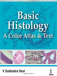
Basic Histology : A Color Atlas & Text
by V Subhadra Devi (Author),
Jaypee Brothers Medical Pub (P) Ltd (Publisher)
Product Specifications
Book Description
This atlas provides undergraduate medical students with an understanding of the
histological structures of various tissues and functional correlation. Beginning
with an introduction to histology, microscopy and tissue preparation for
microscopy, the following chapters illustrate histological aspects of different
tissues (epithelial, connective, muscular and nervous), in different systems of
the body. Each chapter concludes with a table summarising the microscopic
structure of organs in the relevant system, and their function. The final
chapter presents sample histology slides to enhance learning. Highly illustrated
with nearly 340 clinical images and tables, the book also includes multiple
choice and descriptive questions to assist revision. Key points Provides
undergraduate medical students with an
Book Specifications
ISBN-13 9789352501786
Language English
Binding Paper Back
Publisher Jaypee Brothers Medical Pub (P) Ltd
Publishing Date 2016-05-28
Product Edition 1
Total Pages 330